The agent that causes CWD and other TSEs has not been completely characterized. However, the theory supported by most scientists is that TSE diseases are caused by little understood proteins called prions. Prions are a form of protein normally found in the cells of the nervous system and other body tissues. Stanley Prusiner, a Nobel Prize winning neurologist, first described an abnormal form of prion resistant to enzymes that break down normal proteins. These abnormal, protease resistant prions are referred to as PrPres. PrPres have the ability to transform normal prions into this abnormal state. As the disease progresses, PrPres accumulate in the brain and lymphoid tissues (lymph nodes and tonsils). Accumulation of these abnormal PrPres, produce tiny sponge-like holes in the brain that are visible microscopically. The word "spongiform" in TSEs describes the sponge-like condition of brain tissue found in infected animals. As the disease progresses, the affected animal loses its basic physical and mental abilities.
-
CWD was first described clinically as a wasting syndrome in captive deer belonging to Colorado research facilities in 1967. A few years later it was described in a Wyoming research facility.
-
CWD was first determined to be a TSE in 1978 by Dr. Elizabeth Williams of the University of Wyoming.
-
The first cases of CWD in wild deer and elk were diagnosed in 1981 in Colorado and 1985 in Wyoming.
-
Beginning in the 1980s, the distribution of CWD in wild deer and elk in Colorado and Wyoming was determined through surveillance by wildlife agencies in those States. Through their efforts, an endemic area for the disease in wildlife in their States was described. This area includes much of northeastern Colorado and southeastern Wyoming.
-
In 2001, discovery of a positive wild mule deer in neighboring Kimball County, NE, extended the endemic area into southwestern Nebraska.
-
From 1996 to June 2002, CWD was diagnosed in farmed elk herds in Colorado, Kansas, Montana, Nebraska, Oklahoma, South Dakota, and the Canadian Provinces of Alberta and Saskatchewan.
-
From 2000 to June 2002, CWD has also been found in wild deer in northwestern Nebraska, southern New Mexico, southwestern South Dakota, south central Wisconsin, northwestern Colorado, and the Canadian Province of Saskatchewan.
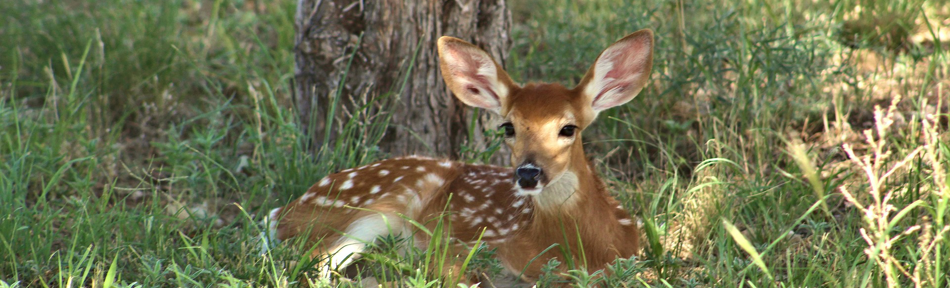
CWD is a slow and progressive disease; signs typically are not seen until the animal is 12-18 months of age and may take as long as 3 or more years. As the disease progresses, deer and elk with CWD show changes in behavior and appearance. These clinical signs may include progressive weight loss, stumbling, tremors, lack of coordination, blank facial expressions, excessive salivation, loss of appetite, excessive thirst and urination, listlessness, teeth grinding, abnormal head posture, and drooping ears. Because of effects on the central nervous system, animals can have difficulty in swallowing, resulting in pneumonia caused by aspiration of food or saliva. Clinical signs of CWD are usually present a few weeks to several months before the animal dies. It should be remembered that many of these signs can be a result of other diseases.
The exact mechanism of transmission is unclear. Evidence suggests CWD is transmitted directly from one animal to another (lateral or horizontal transmission). The route by which the agent is shed from the animal’s body is unknown. However, experimental and circumstantial evidence suggests that indirect transmission from an environment contaminated with the agent appears to be possible. Transmission of CWD has not been associated with any particular feeding practice or regimen in farmed elk or deer. Supplemental feeding of wild elk and deer, however, concentrates the animals and may contribute to disease spread.
A characteristic of all TSE agents is their resistance to conventional disinfectants, high temperatures, and enzymes that normally break down proteins. Recommendations for disinfection of areas in which infected animals have resided are still being developed.
The World Health Organization has reviewed available scientific information and concluded there is no evidence that CWD can be transmitted to humans. Researchers have found no link between the disease and any neurological disease that affects humans including the human TSE disease, Creutzfeldt-Jakob Disease (CJD). Between 1997 and 1998, three cases of sporadic CJD occurred in the U.S. in young adults. These individuals had consumed venison, which led to speculation about possible transmission of CWD from deer or elk to humans. However, review of the clinical records and pathological studies of all three cases by the Centers for Disease Control and Prevention in Atlanta, did not find a causal link to CWD. Ongoing national surveillance for CJD and other neurological cases will remain important for continuing to assess the risk, if any, of CWD transmission to humans.
During the approximately two decades of monitoring, researchers have not found any evidence that CWD can be transmitted to domestic cattle under natural conditions. Ongoing experiments involving oral exposure and contact exposure on heavily CWD contaminated sites have not resulted in infection of cattle. These experiments, however, require additional time before they are completed. CWD has been experimentally transmitted by artificial means to mice, ferrets, mink, goats, squirrel monkeys, and calves.
Currently, CWD is diagnosed by examining brain and lymphoid tissue (lymph nodes and tonsils) from a dead animal. Tests to confirm CWD are performed in a laboratory, using brain tissue. Immunohistochemical (IHC) staining is the most commonly accepted method of detection and is the standard test used by USDA ís National Veterinary Services Laboratories. IHC staining is an antibody-based test. Antibodies bind to abnormal PrPres in the tissue on a slide. Additional steps in the test allow a colored agent to be bound to the abnormal PrPres-antibody complex. Accumulations of color indicate the presence of the abnormal PrPres when the slide is examined microscopically. A CWD-positive animal is one in which the presence of abnormal PrPres has been confirmed in the brain or lymphoid tissues.
A research team in Colorado has recently developed a live animal test for CWD based on the collection of tonsil biopsies for microscopic examination. This test seems to work well in mule deer, but not in elk, and its application may be limited to special circumstances. Scientists are continuing to work on a number of approaches that may provide a rapid postmortem or live animal test for both deer and elk.
Studies on the distribution of abnormal PrPres in CWD affected deer and elk have shown that the obex portion of the brainstem is the first place that the abnormal PrPres can be detected in the brain. As the disease progresses, the abnormal PrPres can be detected in multiple locations and, finally, throughout the brain. Because of this, it is necessary to test the obex to detect CWD in animals that are in the early stages of the disease. It is possible that other parts of the brain may test negative for the presence of disease while the obex would test positive. For whitetail deer and mule deer (but not elk) some lymphoid tissues from the head (tonsils and retropharyngeal lymph nodes) become positive before the obex does, so these tissues will also be useful in surveillance and monitoring efforts in deer.
A negative test is one in which there is no detectable IHC staining of abnormal PrPres. The interpretation of a negative test depends on the species and the tissue tested. In elk, if the obex is negative, the animal is most likely not infected with the CWD agent. There is the possibility, however, that the animal is infected but the disease process is so early that the abnormal PrPres is not detectable with the current IHC test. Similarly, in whitetail deer and mule deer if the obex and/or the lymphoid tissue from the head are IHC negative, the animal is most likely not infected with CWD. There is the possibility, however, that the disease process is so early that the abnormal PrPres is not detectable by the current IHC test.